View other online activities in From DNA to Beer: Harnessing Nature in Medicine & Industry.
Courtesy National Museum of American History
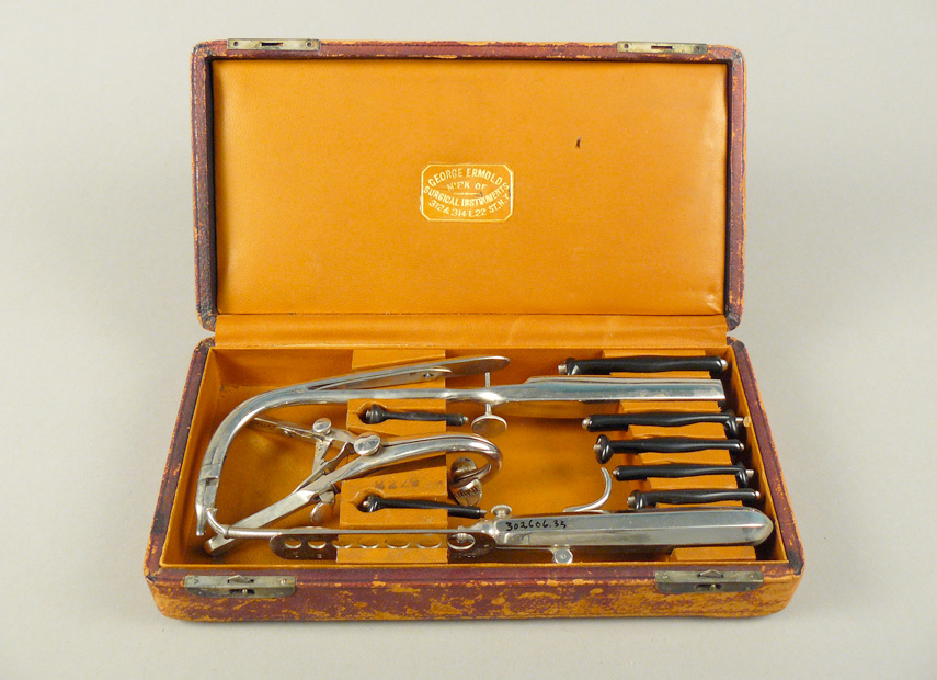
Diphtheria is characterized by a membrane that grows in the throat causing the infected child to struggle for breath. To prevent suffocation, doctors performed tracheotomies and intubation, surgical methods for restoring a breathing passage.
This intubation kit includes a set of seven tubes, sized for children up to age twelve, along with the tools used for inserting and extracting the tubes from the throat.
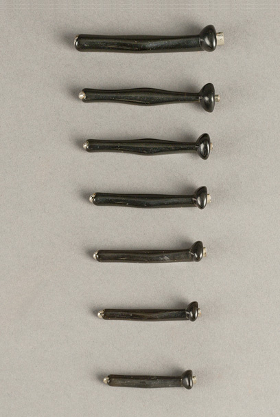
(Left) Hard rubber intubation tubes arranged by length. Courtesy National Museum of American History
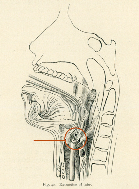
(Right) Cross section of mouth and neck anatomy. Courtesy National Library of Medicine
This kit includes seven hard rubber intubation tubes that vary in their lengths and widths in order to accommodate the windpipe of children up to age 12. The tube has an elliptical, enlarged head and bulging middle, designed to keep it in place in the throat. The illustration shows the placement of the inserted tube in the windpipe.
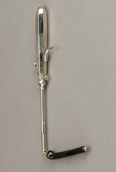
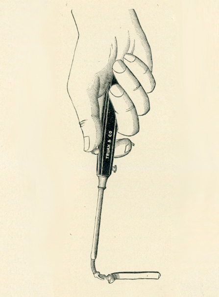
(Left) Metal insertion tool. Courtesy National Museum of American History
(Right) A hand holding an insertion tool. Courtesy National Library of Medicine
The metal insertion tool has a tube holding attachment (bent end) where the black intubation tube is fitted. The long neck of the tool is long enough to reach into the patient’s mouth and insert the tube in the patient’s windpipe.
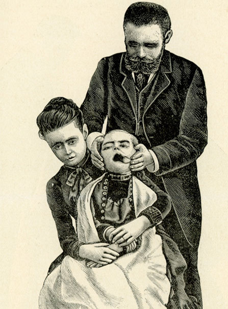
(Left) “Proper position of operator and attendants” from Intubation of the Larynx, Frank E. Waxman, 1888. Courtesy National Museum of American History
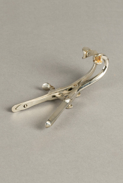
(Right)Metal mouth gag. Courtesy National Library of Medicine
The mouth gag tool keeps the patient’s mouth open for inserting or extracting the intubation tube. The illustration shows how a young patient is held securely by a person, behind whom another individual stands and holds the patient’s mouth open using the gag tool.
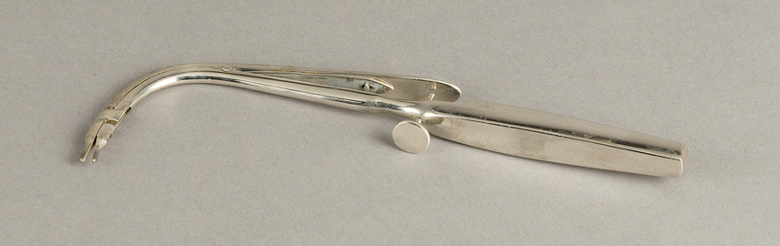
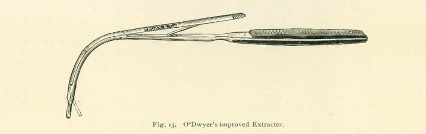
(Top ) L-shaped metal extracting tool. Courtesy National Museum of American History
(Bottom) O’Dwyer’s improved extractor. Courtesy National Library of Medicine
The extraction tool has a pincer end that grabs the intubation tube from the throat before pulling it out of the patient. The illustration shows how the lever by the handle operates the pincer for latching onto the tube.
View other online activities in From DNA to Beer: Harnessing Nature in Medicine & Industry.