Up:
Title Page
Index:
Full Text Index
Contents:
Conference Page
Image Index
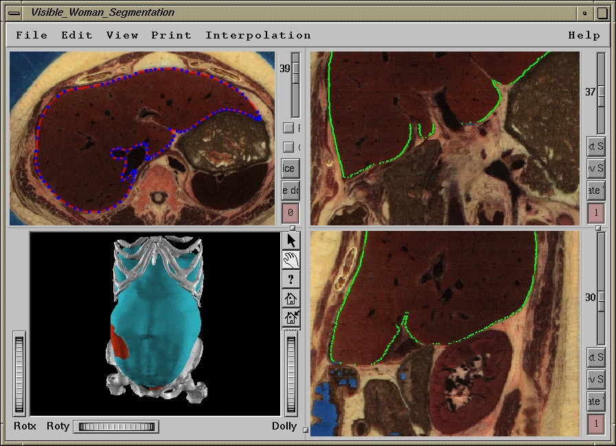
Figure 1. User interface of the segmentation tool.
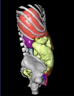
Figure 2 Views of the inner surface of the abdominal cavity.
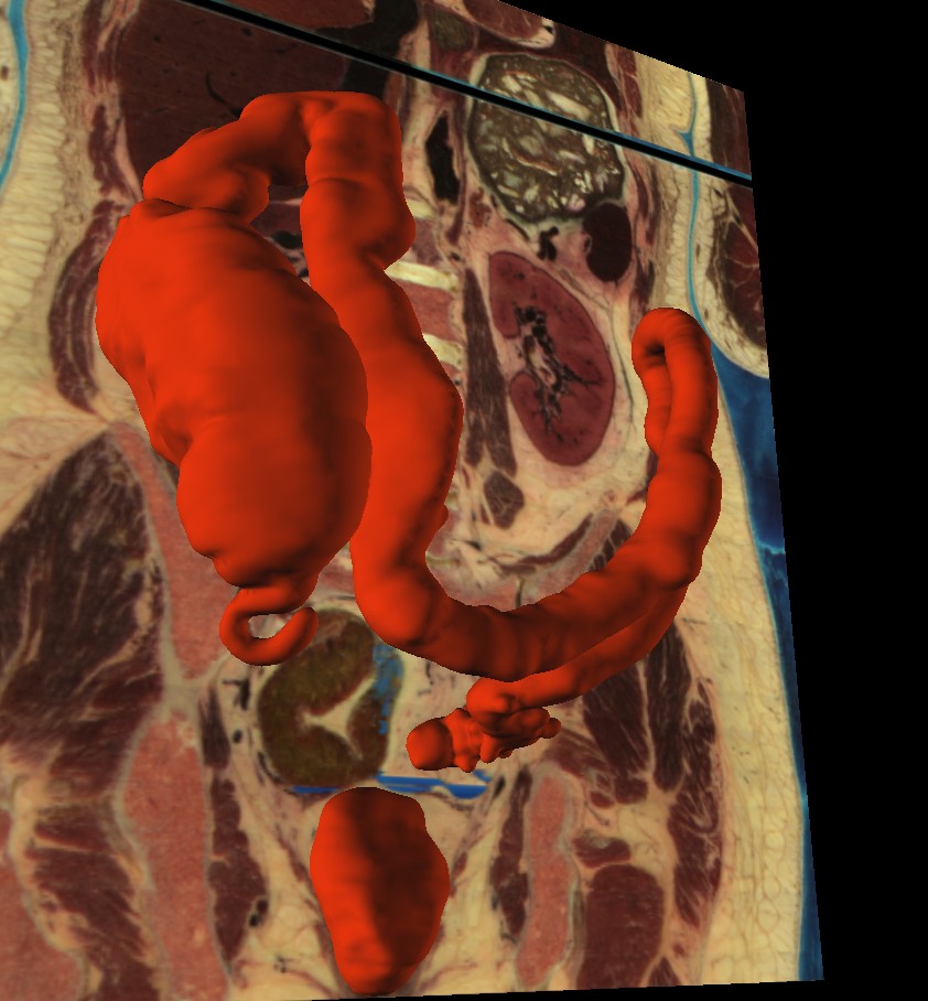
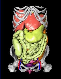
Figure 3. View of the Cecum and the Colon Transversum.
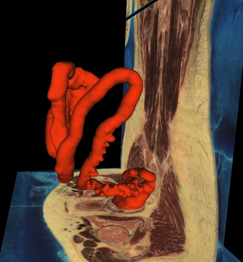
Figure 4. View of the the colon descendens. The detected diverticula are well visible.
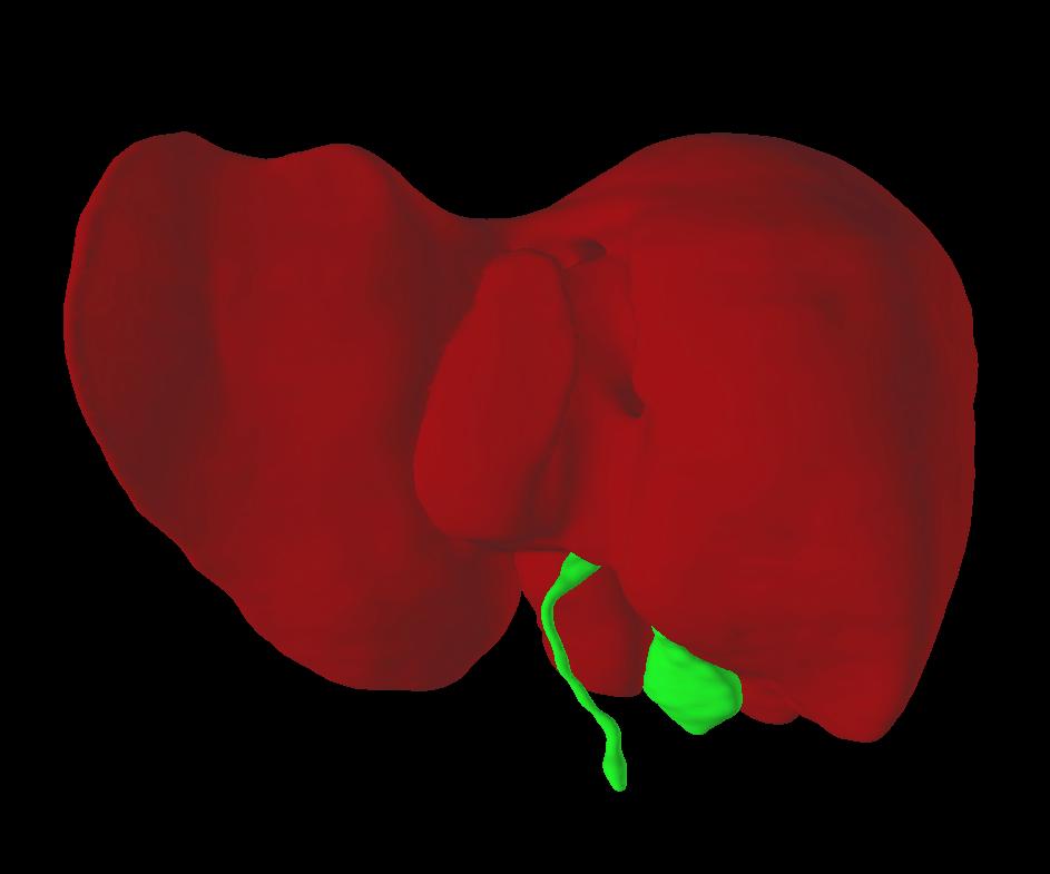
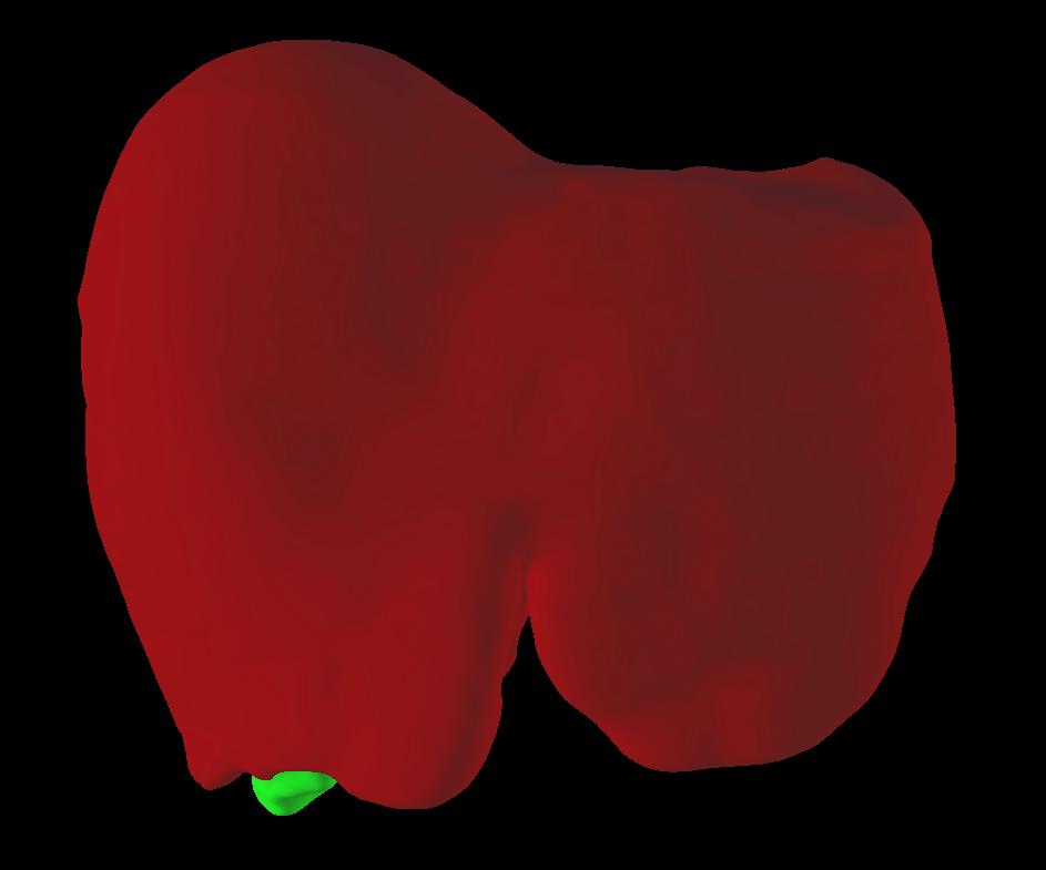
Figure 5. Front and back view of the segmented liver and gallbladder.
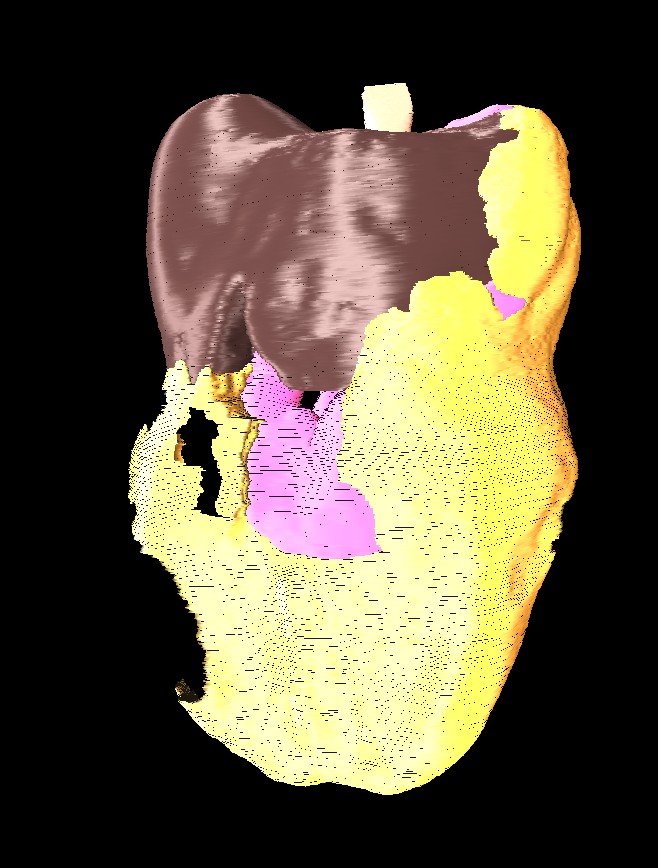
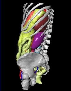
Figure 6 The omentum majus. The stomach becomes visible through the hole ventral of the flexura coli dextra.
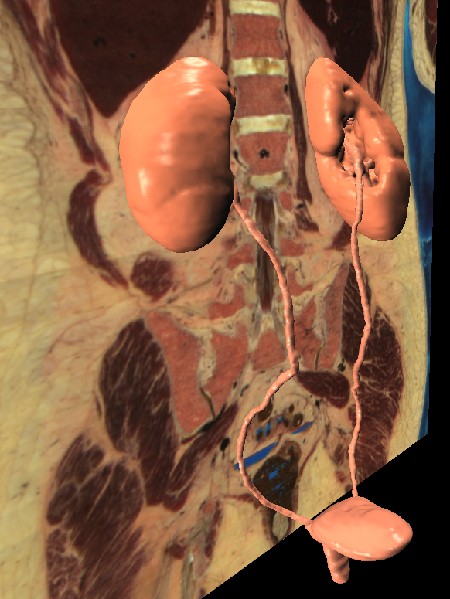
Figure 7. The kidneys and the urinary tract.
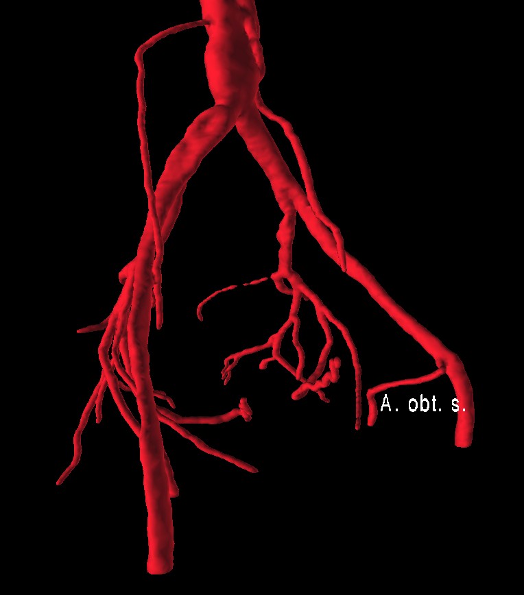
Figure 8. Arteries of the Pelvis minor.
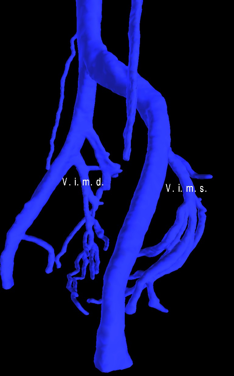
Figure 9. Veins of the Pelvis minor.
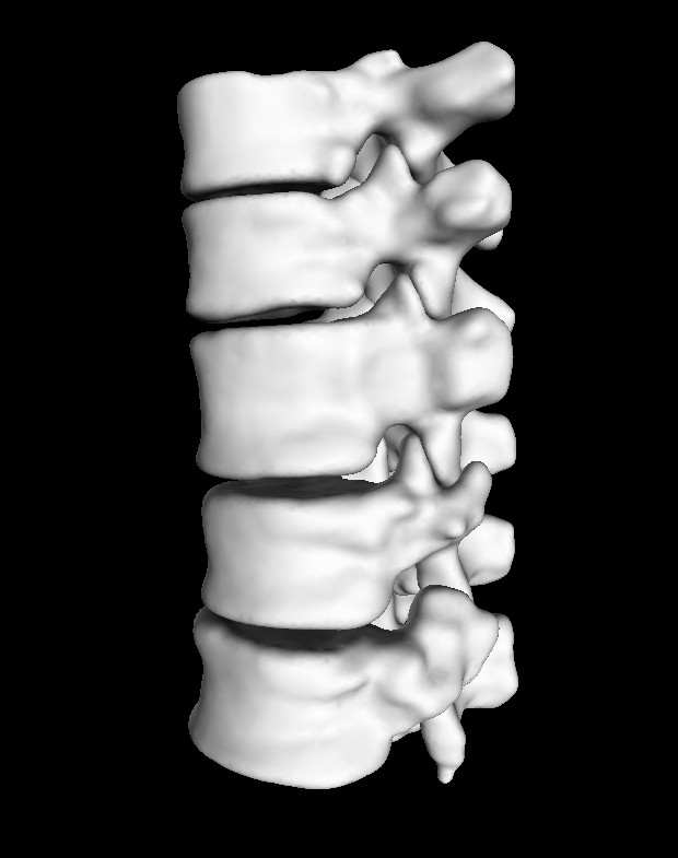
Figure 10. The thoracic vertebrae from VIII to XII. The strong deviation in the height of the Vertebra thoracalis X is well visible.
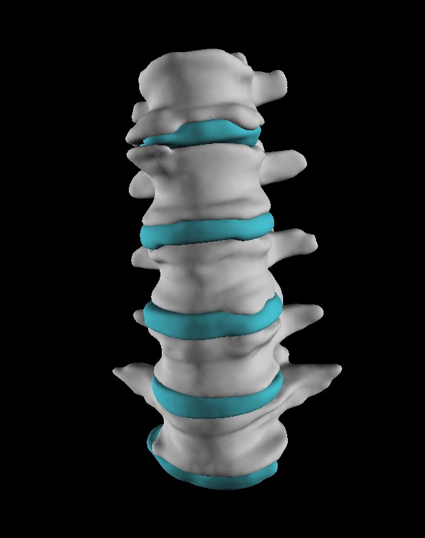
Figure 11. The osteophytic bridge over the discus intervertebralis between vertebrae lumbales I and II.
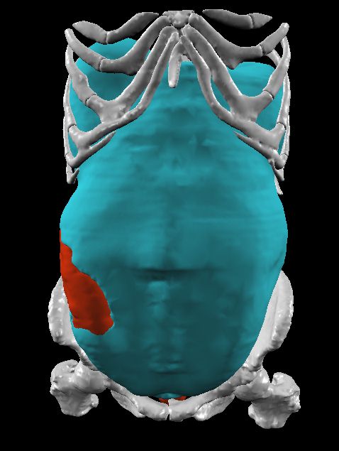
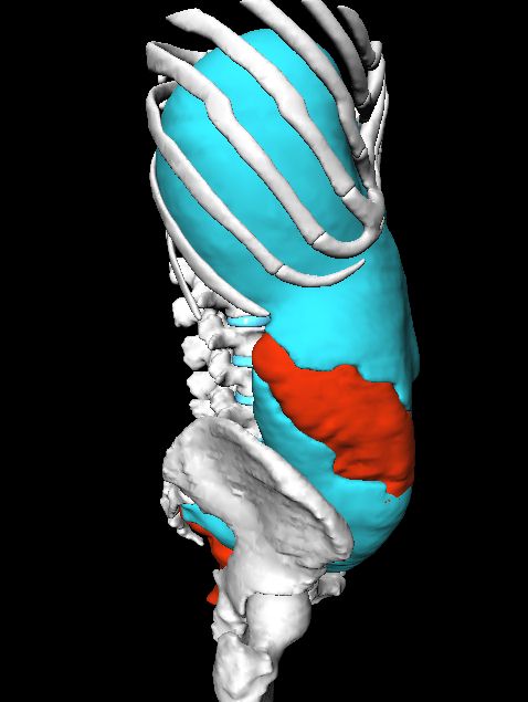
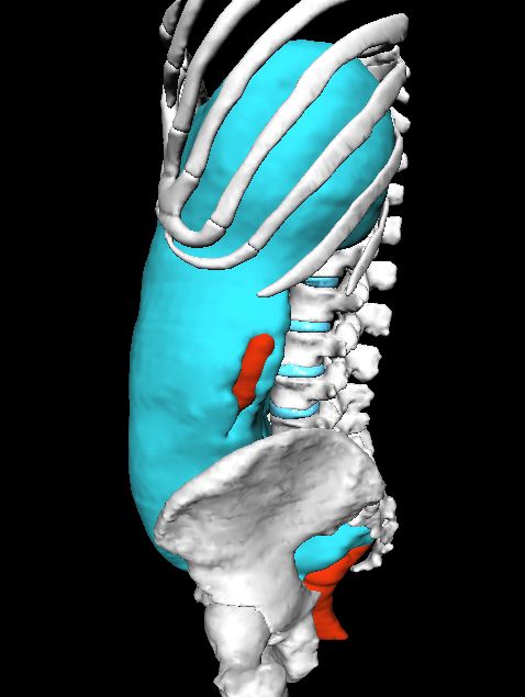
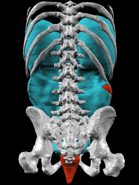
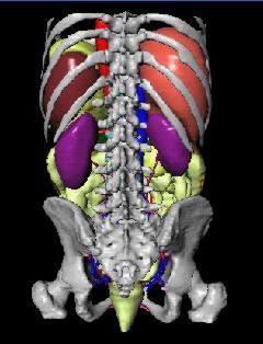
Figure 12. Views of the complete abdominal anatomical model.
Up:
Title Page
Index:
Full Text Index
Contents:
Conference Page

 Figure 10. The thoracic vertebrae from VIII to XII. The strong deviation
in the height of the Vertebra thoracalis X is well visible.
Figure 10. The thoracic vertebrae from VIII to XII. The strong deviation
in the height of the Vertebra thoracalis X is well visible.