Technologies

Skull showing gun shot trauma Male profile, 1950s
Skull showing keyhole gunshot trauma, about 1861-1865
Heart of a 26-year-old man, perforated by a bullet, New York, 1937
Leg bone from the Ragsdale Gunshot Wound Study, 1984
Chest plates commissioned by Frances Glessner Lee, about 1940
Bloodstain, blisters, bullet holes, 1864
Medical professor Johann Ludwig Casper was perhaps the first author to use richly colored lithographs to illustrate forensic pathology. The plates show specific postmortem examinations, some of them experiments on cadavers.
Johann Ludwig Casper, M.D., Atlas zum Handbuch der gerichtlichen Medicin [Atlas for the Manual of Legal Medicine], Berlin; Artist: Hugo Troschel; Lithographer: Winckelmann & Sons
National Library of Medicine
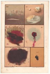
Image 7 of 7

Reading gunshot patterns

The mass manufacture of guns in the 19th century led to an epidemic of gunshot wounds incurred in wars, violent crimes, suicides, and accidents. The study of gunshot wounds became an integral part of criminal investigation and forensic pathology.