Exhibition Images

Previous Images
Bloodstain, blisters, bullet holes, 1864
Head and hand of a drownee, 1864
Decomposed stomach, 1864
Rope marks and upper thigh, 1864
The color of the lungs of dead newborn children: stillborn, newborn who have taken a breath, newborn whose lungs have been artificially inflated, 1864
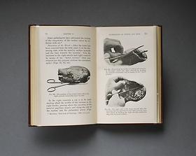
T. N. Kelynack, M.D., The Pathologist's Handbook: A Manual for the Post-Mortem Room, London, 1899
Leg bone from the Ragsdale Gunshot Wound Study, 1984
Chest plates commissioned by Frances Glessner Lee, about 1940
Next Images
T. N. Kelynack, M.D., The Pathologist's Handbook: A Manual for the Post-Mortem Room, London, 1899
Image 24 of 43

Upon a View of the Body

T. N. Kelynack, M.D., The Pathologist's Handbook: A Manual for the Post-Mortem Room, London, 1899
In the late 19th and early 20th centuries, textbooks increasingly used photographs to illustrate forensic pathology and the techniques of postmortem examination. Photographs of this period were often retouched and silhouetted to simplify and highlight the image.
National Library of Medicine