U.S. flag

An official website of the United States government

Official websites use .gov
A .gov website belongs to an official government organization in the United States.

Secure .gov websites use HTTPS
A lock ( ) or https:// means you’ve safely connected to the .gov website. Share sensitive information only on official, secure websites.

History of Medicine

About   |  Collections   |  Exhibitions   |  Research Tools   |  Copyright   |  Get Involved   |  Visit   |  Contact

Banner containing six icons that link to the individual pages of the website.  From left to right: 1. Image of three embryos, that links to the Embryology page.  2. Image of conjoined twins from a 15th century book, that links to the Age of Superstition page. 3. Image of Millie-Christine McCoy, that links to the Marvels on Exhibit page. 4. Image of Clara and Alta Rodriguez, that links to the Separation Surgeries page.  5. Image of a fine engraving of two sets of conjoined twins, that links to the Gallery of Images page.  6. Image of a title page, that links to the bibliography page.   Below the banner, to the left of the links to each page is a large eighteenth century woodcut illustration of female twins joined at the back. From 'Monsters' to Modern Medical Miracles: selected moments in the history of conjoined twins from medieval to modern times. Embryology and Classification of Conjoined Twins Age of Superstition Marvels on Exhibit Separation Surgeries Gallery of Images Bibliography

Embryology of Conjoined Twins

Identical twins develop when a single fertilized egg, also known as a monozygote, splits during the first two weeks of conception. Conjoined twins form when this split occurs after the first two weeks of conception. The monozygote does not fully separate and eventually develops into a conjoined fetus that shares one placenta, one amniotic sac, and one chorionic sac. Because the twins develop from a single egg, they will also be the same sex. The extent of separation and the stage at which it occurs determine the type of conjoined twin, i.e., where and how the twins will be joined.

Nineteenth century drawing of 5 embryos of conjoined twins showing the partially separated germ segments.

The diagram at left illustrates the germ layers in embryos of conjoined twins. The proximity of the segments determines how much shared tissue there will be. The further apart the segments, the greater the likelihood that the organs will develop fully in each fetus. If the segments are at their farthest point, there will only be a minimum of tissue and cartilage joining the twins, i.e. omphalopagus twins will develop.

Classification of Conjoined Twins

Isidore Geoffroy Saint-Hillaire was the first teratologist to classify conjoined twins, using Greek etymology to describe the twins in terms of their shared anatomy. Many of his terms are still in use today. Listed below are some commonly occurring types of conjoined twins, examples of which may be viewed throughout this exhibit.

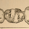
Nineteenth century drawing of twins joined at the crowns of their heads.

Craniopagus: joined at the cranium (head).

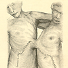
Nineteenth century drawing of adult male omphalopagus or xiphopagus twins.  The twins share a band of tissue in the region of the umbilicus.

Omphalopagus or xiphopagus: joined in the region of the umbilicus.

Late fifteenth century woodcut illustration of female twins,  joined at the chest.

Thoracopagus: joined at the thoracic cavity (chest).

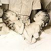
Twentieth century photograph of newborn ischiopagus twin girls being held by a nurse.  The twins are joined at the inferior margins of the coccyx and sacrum and have separate spinal columns.

Ischiopagus: joined at the inferior margins of the coccyx and sacrum, with two separate spinal columns.

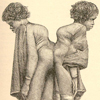
Nineteenth century drawing of adult female African-American pygopagus twins,  joined at the lateral and posterior surfaces of the coccyx and sacrum.

Pygopagus: joined at the lateral and posterior surfaces of the coccyx and sacrum.

Last Reviewed: March 11, 2024