 Biomedical Research
Current research
Following World War II, federal support for biomedical research was greatly expanded and so was the role of the National Institute of Health, which was renamed the National Institutes of Health to reflect the growth of research functions. Today NIH encompasses seventeen research institutes, two research divisions, the world's largest research hospital, the National Library of Medicine, the National Center for Human Genome Research, the National Center for Research Resources, and the Fogarty International Center. Its operating budget in 1990 was 7.6 billion dollars.
NIH conducts research on its Bethesda, Maryland campus and supports research throughout the United States by means of a competitive grant system. This research deals with every aspect of human biology and almost every disease and disability.
Some of the major topics of research today include: oncogenes and the question of how a normal cell becomes a cancer; the workings of immune cells and how they help to defend the body against cancer; the isolation of genes, including those for blood clotting factors; brain changes in Alzheimer's disease; the application of genetic engineering techniques to vaccine production; the discovery and isolation of bone growth factors; the study of neurotransmitters and modulation in the brain; cancer risk from passive smoking; new uses of the laser for the treatment of eye diseases; the construction of artificial chromosomes; the development and refinement of new body imaging technologies, such as magnetic resonance imaging (MRI) and, of course, the study of AIDS.
Gallery
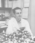
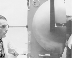
In 1968, Dr. Marshall W. Nirenberg of the National Heart, Lung, and Blood Institute became the first of four NIH Nobel laureates to date. He won the Nobel Prize in Physiology or Medicine for his work in translating the genetic code and its function in protein synthesis; he is shown here with his molecular models.
c. 1965
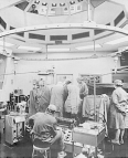
The National Heart, Lung, and Blood Institute has been a leader in conducting and supporting clinical studies of heart disease, including open heart surgery and artificial hearts, as well as educating the public about the prevention of heart attacks through control of high blood pressure, control of high levels of blood cholesterol, and cessation of smoking. Research advances and lifestyle changes have helped lower the death rate from heart disease by 43 percent since 1972.
c. 1970
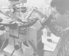
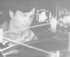
Researcher at the National Heart, Lung, and Blood Institute's Laboratory of Kidney and Electrolyte Metabolism uses fluorescence microscopy to study kidney physiology, specifically kidney tubule transport. Other important research work on kidney function, such as improving the prevention and management of end-stage renal disease and improving dialysis techniques, is done at the National Institute of Diabetes and Digestive and Kidney Diseases.
c. 1987
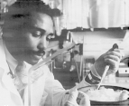
Researcher at the National Heart, Lung, and Blood Institute engaged in the study of sickle cell anemia, a genetic blood disease which, in the United States, affects primarily Afro-Americans and is caused by an abnormal hemoglobin molecule. A new test, known as chorionic villus biopsy, promises to advance the prenatal diagnosis of genetic blood diseases, including sickle cell anemia, from the second trimester of pregnancy to the first. Tissue from the chorionic villi, hairlike projections of the membrane that surrounds the early embryo, can be removed before the 10th week of pregnancy and analyzed immediately for chromosomal or biochemical defects.
c. 1987
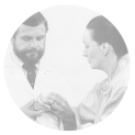
The National Institute of Diabetes and Digestive and Kidney Diseases (NIDDK) has pioneered in the study and treatment of diabetes, the fifth leading cause of death in the United States. The NIDDK supported research which led to the development of the insulin pump. A physician instructs a patient in its use.
c. 1987
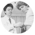
Dr. John Klippel of the National Institute of Arthritis and Musculoskeletal and Skin Diseases (NIAMS) examines and measures the extension of the fingers of a patient with lupus, a disease characterized by eruption of scarred lesions and inflamed joints. Researchers at the NIAMS elaborated the underlying immunologic mechanisms in systemic lupus erythematosus (SLE), developed more effective treatments, and dramatically improved survival.
c. 1987
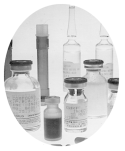
The development and testing of new and promising experimental drugs or vaccines for a variety of ailments is one of the key functions of the National Institutes of Health researchers.
c. 1987
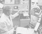
Increasingly sophisticated instrumentation such as this fast atomic bombardment mass spectrometer, which separates chemical components, is being used at the FDA's Center for Biologics Evaluation and Research to determine the purity and potency of biologics and other products.
c. 1987
Laser technology is being applied to biomedical research by scientists at the FDA's Center for Biologics Evaluation and Research. Here a laser cell separation machine is separating a line of cells, such as T-cells, for immunology studies.
c. 1987
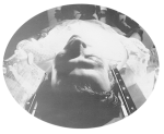
A computed tomography (CT) scan, a noninvasive method of getting good cross-sectional images of the body, is being done on a patient at the NIH's Warren U. Magnuson Clinical Center. The Clinical Center is the world's largest hospital devoted solely to biomedical research where physicians from all the different NIH institutes, together with the Center's staff, pursue clinical and laboratory studies related to patient care.
c. 1987
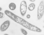
Legionnaires' disease was first recognized in July 1976, when a sudden outbreak of pneumonia, resulting in several deaths, occurred mostly in persons who had attended an American Legion convention in Philadelphia. Researchers Charles C. Shepard (1914-85) and Joseph E. McDade of the Centers for Disease Control and Prevention were the first in 1977 to identify the disease-causing bacterium -- Legionella pneumophilis, which is pictured here. Since then more than 20 species in the Legionella genus have been identified and the mystery surrounding many illnesses associated with them solved, in the grand tradition of the microbe hunters of the late 19th and early 20th centuries. Many of these modern day microbe hunters or epidemiologists were trained in the Centers for Disease Control and Prevention's Epidemic Intelligence Service, which was established by Dr. Alexander Laugmuir in 1951.
c. 1977
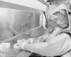
Researchers, such as this man in the Centers for Disease Control and Prevention's new maximum containment virology laboratory, use the most advanced technology available to study dangerous organisms like the Lassa, Machupo, Ebola, and AIDS viruses that cause deadly diseases for which no cure or vaccine exists. Statistics about these and other diseases are published in the Morbidity and Mortality Weekly Report (MMWR), a widely read publication around the world. Weekly reporting of morbidity and mortality statistics to the Public Health Service began in 1893. Various bureaus of the Service have published these reports. Since 1961, the Center for Disease Control and Prevention's Epidemiology Program Office has been responsible for publishing the MMWR.
c. 1987
Researcher at the National Eye Institute (NEI) tests the peripheral and central visual fields of a patient to find areas of visual loss. The NEI has helped pioneer the use of lasers in preventing visual loss from diabetes and other eye diseases, and is testing new medicines that may he able to prevent diabetes-induced damage to the retina and other tissues.
c. 1987
Researcher at the National Institute of Dental Research (NIDR) uses computers to measure very small changes in teeth and their surrounding tissues that are not detectable with conventional dental X-rays. Since its creation in 1948, the NIDR has been the primary sponsor of dental research in the United States. Much of its work was focused on preventing tooth decay, through such programs as community water fluoridation.
c. 1987
Dr. Robert C. Gallo, since 1972 chief of the National Cancer Institute's Laboratory of Tumor Cell Biology, is an internationally prominent investigator of human viruses and tumor cells. He played a leading role in isolating and characterizing the family of human viruses to which the AIDS causing virus, HIV (human immunodeficiency virus), belongs. Gallo and his colleagues are also responsible for the development of a blood test to detect HIV antibodies in blood collected for transfusions.
1987
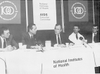
Vice-President George Bush addresses the AIDS Executive Committee during the centennial year of the National Institutes of Health. As the NIH enters its second century it faces one of its greatest research challenges -- a cure and vaccine against the deadly viral disease AIDS (acquired immunodeficiency syndrome), which has become the major scourge of the late twentieth century. To the left of the Vice-President is Dr. James B. Wyngaarden, director of the NIH, and to the right of the Vice-President is Dr. Anthony Fauci, director of the NIH's National Institute of Allergy and Infectious Diseases and coordinator of NIH research on AIDS.
1987
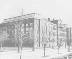
The National Library of Medicine was established in 1836 as the Library of the Army Surgeon General's Office. The Armed Forces Institute of Pathology and its Medical Museum were founded in 1862 as the Army Medical Museum. Throughout their history the Army Medical Library and the Army Medical Museum often shared quarters. This red-brick building was built on the Mall in Washington, D.C., in 1887 for these two institutions. It was torn down in the late 1960s to make room for the Hirshorn Museum of Art. By an act of Congress in 1936 the Library collection was transferred from the Department of Defense to the Public Health Service of the Department of Health, Education and Welfare and renamed the National Library of Medicine. The Library moved to its current quarters in Bethesda, Maryland, on the campus of the National Institutes of Health in 1962.
c. 1890
Under the very able leadership of Dr. John Shaw Billings (1838-1913), a Civil War surgeon who served as director from 1865 to 1895, the Library increased in size from about 2,000 volumes to over 100,000 volumes of books and bound serials and expanded from serving primarily military medical officers to serving all physicians. It soon became the leading medical library in the United States and then the world. The first issue of Index Medicus, a comprehensive monthly medical bibliography with a subject and author index to articles published in medical journals around the world, was published in 1879.
c. 1950
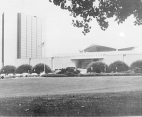
The National Library of Medicine is now the world's largest medical research library. Its holdings include more than 3.5 million books, journals, technical reports, theses, pamphlets, photographs, manuscripts and audiovisual materials covering more than 40 biomedical areas and related subjects. The Library also houses one of the world's finest historical collections of rare medical texts and manuscripts. The 10-story Lister Hill Center was built in 1980 to house the Library's expanding computer facilities, audiovisual studios, and research and development laboratories. With materials in 70 languages and international exchange capabilities the Library can serve health professionals worldwide.
c. 1983
In order to keep up with the ever-increasing volume of biomedical literature that had to be included in the Index Medicus bibliography, the Library turned to computers in the early 1960s. An extensive computerized literature retrieval system, known as MEDLARS, became totally operational in 1964. It performed thousands of searches before on-line searching capabilities and data bases, such as MEDLINE, became available in 1971. Through MEDLINE, health professionals and other interested individuals have immediate access to more than 3 million journal article references accumulated since 1965 and growing at a rate of over 300,000 a year.
c. 1964


